
Page 272 - Vitamin D and Cancer
P. 272
11 Vitamin D and Hematologic Malignancies 259
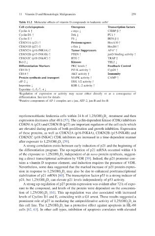
Table 11.2 Molecular effects of vitamin D compounds in leukemic cells a
Cell cycle/apoptosis Oncogenes Transcription factors
Cyclin A ↑ c-myc ↓ C/EBP b ↑
Cyclin D1 ↑ Dek ↓ PU.l ↑
Cyclin E ↑ Fli ↓ IRF8 b ↑
CDKN1A (p21) ↑ Protooncogenes HoxA10 ↑
CDKN1B (p27) ↑ c-fms ↓ HoxB4 ↑
CDKN2A (p16-INK4A) ↑ Tumor Suppressors AP-l ↑
b
CDKN2B (p15-INK4B) ↑ PTEN ↑ junD binding activity ↑
CDKN2C (p18-INK4C) ↑ BTG ↑ TRAP ↑
Bcl-2 ↓ Kinases TEL2 ↓
Differentiation Markers PKC levels ↑ Feedback Control
CD11b ↑ PI3-K activity ↑ Cyp24 ↑
CD14 ↑ AKT activity ↑ Immunity
Protein synthesis and transport MAPK activity ↑ CAMP ↑
eIF-2 ↓ ERK 1/2 activity ↑
Importins ↓ KSR-1,-2 activity ↑
Exportins -1,-5,-7, -t ↓
a Regulation of expression or activity may occur either directly or as a consequence of
differentiation. See text for details
b Putative components of AP-1 complex are c-jun, ATF-2, jun-B and fos-B
myelomonoblastic leukemia cells within 24 h of 1,25(OH) D -treatment and then
2
3
expression decreases after 48 h [57]. The cyclin-dependent kinase (CDK) inhibitors
CDKN1A (p21) and CDKN1B (p27) are important regulators of the cell cycle which
are elevated during periods of both proliferation and growth inhibition. Expression
of these proteins, as well as CDKN2A (p16-INK4A), CDKN2B (p15-INK4B) and
CDKN2C (p18-INK4C) CDK inhibitors are increased in a time-dependent manner
after exposure to 1,25(OH) D [59].
3
2
A strong correlation exists between early induction of p21 and the beginning of
the differentiation program. The up-regulation of p21 mRNA occurred within 4 h
of the exposure to 1,25(OH) D independent of de novo protein synthesis, suggest-
3
2
ing a direct transcriptional activation by VDR [59]. Indeed, the p21 promoter con-
tains a vitamin D response element, and induction requires the presence of VDR.
Nevertheless, some data suggested that the marked increase of p21 protein expres-
sion in response to 1,25(OH) D may also be due to enhanced posttranscriptional
3
2
stabilization of p21 mRNA [60]. The transcription factor p53 is a strong inducer of
p21; but 1,25(OH) D can elevate p21 levels independently of p53 activity.
3
2
A strong up-regulation of p27 protein expression was evident after 72 h of expo-
sure to the compound, and levels of the protein were dependent on the concentra-
tion of 1,25(OH) D [61]. This up-regulation was also associated with increased
3
2
levels of Cyclins D1 and E, coinciding with a G1 arrest. These results suggested a
prominent role of p27 in mediating the antiproliferative activity of 1,25(OH) D in
3
2
this cell line. The 1,25(OH) D has a protective effect against apoptosis in HL-60
2
3
cells [62, 63]. In other cell types, inhibition of apoptosis correlates with elevated