
Page 160 - Vitamin D and Cancer
P. 160
7 Induction of Differentiation in Cancer Cells by Vitamin D 147
have been identified, and these include changes in “transepithelial electrical resistance”
and ubiquitin, as based on matrix-assisted laser desorption/ionization time-of-flight
mass spectrometry (MALDI-TOFMS). The latter procedure generates specific mass
spectral fingerprints characteristics of cell differentiation, and it was suggested that
ubiquitin can be a marker of differentiation of the T84 human colon carcinoma cell
line [28]. In another colon cancer cell line, SW80, 1,25D was shown to induce easily
recognizable morphological changes indicative of differentiated epithelial-like phe-
notype [29]. These morphological changes include consequences of the adherence
to the culture substratum, which make the cells look flat and polygonal, and it was
demonstrated that these cells have reduced tumorigenicity when implanted into
athymic mice. Thus, the epidemiological data which indicate that 1,25D has a nega-
tive effect on the incidence of human colorectal cancer [30, 31] are well supported
by the in vitro studies of 1,25D-induced differentiation of colon carcinoma cell lines.
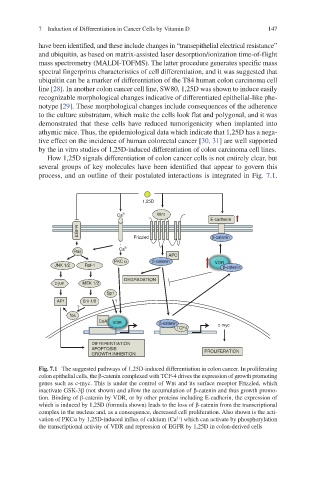
How 1,25D signals differentiation of colon cancer cells is not entirely clear, but
several groups of key molecules have been identified that appear to govern this
process, and an outline of their postulated interactions is integrated in Fig. 7.1.
1,25D
Ca 2+ Wnt
E-cadherin
EGFR Frizzled β-catenin
Ras Ca 2+
APC
PKC α β-catenin VDR
JNK 1/2 Raf-1 β-catenin
DEGRADATION
c-jun MEK 1/2
Sp1
AP1 Erk 1/2 ?
?
fos
CoA VDR β-catenin
TCF4 c-myc
DIFFERENTIATION
APOPTOSIS PROLIFERATION
GROWTH INHIBITION
Fig. 7.1 The suggested pathways of 1,25D-induced differentiation in colon cancer. In proliferating
colon epithelial cells, the b-catenin complexed with TCF-4 drives the expression of growth promoting
genes such as c-myc. This is under the control of Wnt and its surface receptor Frizzled, which
inactivate GSK-3b (not shown) and allow the accumulation of b-catenin and thus growth promo-
tion. Binding of b-catenin by VDR, or by other proteins including E-cadherin, the expression of
which is induced by 1,25D (formula shown) leads to the loss of b-catenin from the transcriptional
complex in the nucleus and, as a consequence, decreased cell proliferation. Also shown is the acti-
vation of PKCa by 1,25D-induced influx of calcium (Ca ) which can activate by phosphorylation
2+
the transcriptional activity of VDR and repression of EGFR by 1,25D in colon-derived cells