
Page 341 - Vitamin D and Cancer
P. 341
328 B.W. Hollis
15.1 Introduction
In 1971, Haddad and Chyu published a seminal paper in The Journal of Clinical
Endocrinology and Metabolism that described a competitive protein-binding assay
(CPBA) for the determination of circulating 25-hydroxycalfierol [25(OH)D] in
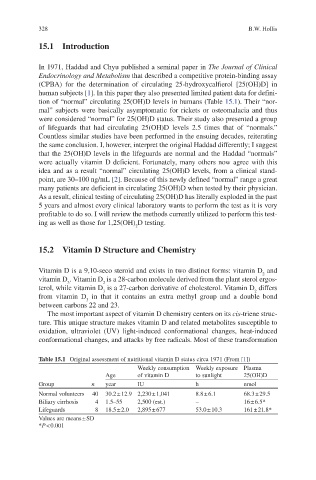
human subjects [1]. In this paper they also presented limited patient data for defini-
tion of “normal” circulating 25(OH)D levels in humans (Table 15.1). Their “nor-
mal” subjects were basically asymptomatic for rickets or osteomalacia and thus
were considered “normal” for 25(OH)D status. Their study also presented a group
of lifeguards that had circulating 25(OH)D levels 2.5 times that of “normals.”
Countless similar studies have been performed in the ensuing decades, reiterating
the same conclusion. I, however, interpret the original Haddad differently; I suggest
that the 25(OH)D levels in the lifeguards are normal and the Haddad “normals”
were actually vitamin D deficient. Fortunately, many others now agree with this
idea and as a result “normal” circulating 25(OH)D levels, from a clinical stand-
point, are 30–100 ng/mL [2]. Because of this newly defined “normal” range a great
many patients are deficient in circulating 25(OH)D when tested by their physician.
As a result, clinical testing of circulating 25(OH)D has literally exploded in the past
5 years and almost every clinical laboratory wants to perform the test as it is very
profitable to do so. I will review the methods currently utilized to perform this test-
ing as well as those for 1,25(OH) D testing.
2
15.2 Vitamin D Structure and Chemistry
Vitamin D is a 9,10-seco steroid and exists in two distinct forms: vitamin D and
2
vitamin D . Vitamin D is a 28-carbon molecule derived from the plant sterol ergos-
2
3
terol, while vitamin D is a 27-carbon derivative of cholesterol. Vitamin D differs
2
3
from vitamin D in that it contains an extra methyl group and a double bond
3
between carbons 22 and 23.
The most important aspect of vitamin D chemistry centers on its cis-triene struc-
ture. This unique structure makes vitamin D and related metabolites susceptible to
oxidation, ultraviolet (UV) light-induced conformational changes, heat-induced
conformational changes, and attacks by free radicals. Most of these transformation
Table 15.1 Original assessment of nutritional vitamin D status circa 1971 (From [1])
Weekly consumption Weekly exposure Plasma
Age of vitamin D to sunlight 25(OH)D
Group n year IU h nmol
Normal volunteers 40 30.2 ± 12.9 2,230 ± 1,041 8.8 ± 6.1 68.3 ± 29.5
Biliary cirrhosis 4 1.5–55 2,500 (est.) – 16 ± 6.5*
Lifeguards 8 18.5 ± 2.0 2,895 ± 677 53.0 ± 10.3 161 ± 21.8*
Values are means ± SD
*P < 0.001