
Page 209 - Vitamin D and Cancer
P. 209
196 F.S.G. Cheung and J.K.V. Reichardt
D , either ingested or synthesized enter the liver where they are metabolized by liver
3
mitochondrial and microsomal 25-hydroxylase (25-OHase), the gene product of
CYP27A1, to 25OHD . This is the main circulating form of vitamin D [116]. Further
3 3
hydroxylations occur in the proximal tubules of the kidneys where 1,25(OH) D (cal-
2 3
citriol) is produced via kidney 1a-hydroxylase (1a-OHase), the gene product of
CYP27B1 (Fig. 9.1a). It has been also shown that the entire pathway to forming
1,25(OH) D from 7-dehydrocholesterol can occur in the human skin [93, 104], dem-
2 3
onstrating the importance of the human skin in the synthesis of vitamin D.
The 1,25(OH) D produced in the kidney is then transported in the blood and is
2 3
mostly bound to the vitamin D binding protein with only a very small amount of its
free form being able to elicit a biological response [116].
Serum level of 1,25(OH) D is regulated by 25-hydrodxyvitamin D 24-hydroxylase
2 3
(24-OHase) which is encoded by the CYP24A1 gene. The CYP24A1 gene is strongly
induced by 1,25(OH) D [118]. With adequate levels of 1,25(OH) D the 24-OHase
2 3 2 3,
acts on 25OHD and 1,25(OH) D to form the inactive metabolites 24,25(OH) D and
3 2 3 2 3
1a,24,25(OH) D . The expression of CYP27B1 is also down regulated by its own
2 3
gene product 1,25(OH) D [109]. Thus by inducing CYP24A1 and down regulating
2 3
CYP27B1, 1,25(OH) D possesses its own feedback regulation via these two genes.
2 3
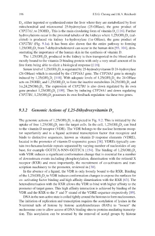
9.3.2 Genomic Actions of 1,25-Dihydroxyvitamin D
3
The genomic actions of 1,25(OH) D is depicted in Fig. 9.2. This is initiated by the
3
2
uptake of free 1,25(OH) D into the target cells. In the cell, 1,25(OH) D can bind
3
2
3
2
to the vitamin D receptor (VDR). The VDR belongs to the nuclear hormone recep-
tor superfamily and is a ligand activated transcription factor that recognize and
binds to distinctive sequences, known as vitamin D response elements (VDRE),
located in the promoter of vitamin D responsive genes [38]. VDREs typically con-
tain two hexanucleotide repeats separated by varying number of nucleotides of any
base, for example GGTTCA-NNN-GGTTCA [154]. The binding of 1,25(OH) D
2
3
with VDR induces a significant conformation change that is essential for a number
of downstream events including phosphorylation, dimerisation with the retinoid X
receptor (RXR) and most importantly, the recruitment of co-activators and tran-
scription machinery to the promoter, reviewed in [38].
In the absence of a ligand, the VDR is only loosely bound to the RXR. Binding
of the 1,25(OH) D to VDR induces conformation changes to expose the surfaces for
3
2
co- activating factor binding and high affinity dimerization with the RXR [63]. The
heterodimerisation with the RXR allows the VDR to bind with higher affinity to the
promoter of target genes. This high affinity interaction is achieved by binding of the
VDR and the RXR to the 3¢ and 5¢ strand of the VDRE sequence respectively [89].
DNA in the non-active state is coiled tightly around the histones to form nucleosomes.
The initiation of replication and transcription requires the acetylation of lysines in the
N-terminal tails of histone by histone acetyltransferases (HATs) to “loosen” the
nucleosome core to allow access of DNA binding sites to proteins mediating transcrip-
tion. This acetylation can be reversed by the removal of acetyl groups by histone