Page 299 - Vitamin D and Cancer
P. 299
286                                                   G.M. Zinser et al.


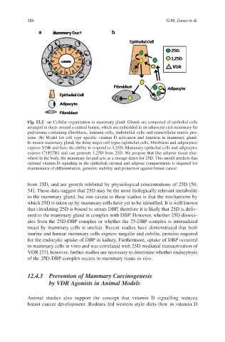
            Fig. 12.2  (a) Cellular organization in mammary gland. Glands are composed of epithelial cells
            arranged in ducts around a central lumen, which are embedded in an adipocyte rich mammary fat
            pad/stroma containing fibroblasts, immune cells, endothelial cells and extracellular matrix pro-
            teins.  (b)  Model  for  cell  type  specific  vitamin  D  activation  and  function  in  mammary  gland.
            In mouse mammary gland, the three major cell types (epithelial cells, fibroblasts and adipocytes)
            express VDR and have the ability to respond to 1,25D. Mammary epithelial cells and adipocytes
            express CYP27B1 and can generate 1,25D from 25D. We propose that like adipose tissue else-
            where in the body, the mammary fat pad acts as a storage depot for 25D. This model predicts that
            optimal vitamin D signaling in the epithelial, stromal and adipose compartments is required for
            maintenance of differentiation, genomic stability and protection against breast cancer


            from 25D, and are growth inhibited by physiological concentrations of 25D [50,
            54]. These data suggest that 25D may be the most biologically relevant metabolite
            in the mammary gland, but one caveat to these studies is that the mechanisms by
            which 25D is taken up by mammary cells have yet to be identified. It is well known
            that circulating 25D is bound to serum DBP, therefore it is likely that 25D is deliv-
            ered to the mammary gland in complex with DBP. However, whether 25D dissoci-
            ates from the 25D-DBP complex or whether the 25-DBP complex is internalized
            intact by mammary cells is unclear. Recent studies have demonstrated that both
            murine and human mammary cells express megalin and cubilin, proteins required
            for the endocytic uptake of DBP in kidney. Furthermore, uptake of DBP occurred
            in mammary cells in vitro and was correlated with 25D mediated transactivation of
            VDR [55], however, further studies are necessary to determine whether endocytosis
            of the 25D-DBP complex occurs in mammary tissue in vivo.


            12.4.3   Prevention of Mammary Carcinogenesis
                    by VDR Agonists in Animal Models


            Animal  studies  also  support  the  concept  that  vitamin  D  signalling  reduces
            breast cancer development. Rodents fed western style diets (low in vitamin D

